The development of non-destructive methods for early detection and understanding the mechanism of cold stress in plants is one of the very important aspects of crop breeding programs to which aquaphotomics research aims to contribute with new insights and solutions.
After many years of dedicated research in this area, we are proud to report that the first step towards this goal has been achieved. Our paper “Aquaphotomics Research of Cold Stress in Soybean Cultivars with Different Stress Tolerance Ability: Early Detection of Cold Stress Response” has been published this month in the Special Issue of Molecules journal, Aquaphotomics – Exploring Water Molecular Systems in Nature.
This paper is a product of joint efforts of our well known aquaphotomics team, Assist. Prof. Dr J. Muncan and Prof. Dr R. Tsenkova, and also pioneers of exploration of plant stress using aquaphotomics, Prof. Dr B.M.S. Jinendra and Prof. Dr S. Kuroki. Prof. Jinendra and Prof. Tsenkova authored the first aquaphotomics publication about early diagnosis of mosaic virus infection in soybean using aquaphotomics, more than 10 years ago, paving the way towards many other contributions in biotic stress diagnosis. Prof. Kuroki on the other hand, co-authored the first aquaphotomics paper which reported that the ability of the resurrection plants to survive very long periods without water are directly related to the water self-organization expressed as dynamic changes of water molecular structures in their leaves.
The newly published paper shows that with aquaphotomics, we were able to detect the effects of even very mild cold stress in soybean plants, in a completely non-destructive manner, and further on to even observe that soybean cultivars with different stress tolerance abilities could be distinguished. This paper is only the first one in the planed series of 3 papers, all of which explore the cold stress in soybean from different aspects, but in the future publications we will show how the genotypic differences are actually related to water molecular structure of the leaves resulting in different susceptibility of soybean cultivars to cold stress.
Stay with us and wait for more exciting discoveries in our next soybean publications.
Until then, we hope you will enjoy our first paper in this series and the incredible simplicity of early cold stress detection.
Figure 1. Aquagrams showing differences in the average spectral pattern of all soybean cultivars in the conditions of no stress and during cold stress. (Source: Muncan, J.; Jinendra, B.M.S.; Kuroki, S.; Tsenkova, R. Aquaphotomics Research of Cold Stress in Soybean Cultivars with Different Stress Tolerance Ability: Early Detection of Cold Stress Response. Molecules 2022, 27, 744. doi.org/10.3390/molecules27030744)
Recording of Christmas Webinars are now available on the Aquaphotomics YouTube Channel!
Thank you for joining us at the webinars!
We are in process of planning more upcoming webinars. We will be sure to update everyone when we are ready to go. Thank you for your continued support in Aquaphotomics!
Webinar #1
Webinar #2
Dear Friends and Colleagues,
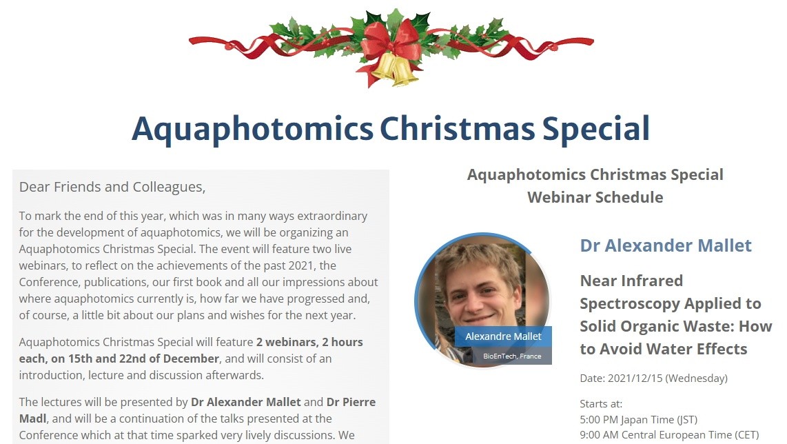
To mark the end of this year, which was in many ways extraordinary for the development of aquaphotomics, we will be organizing an Aquaphotomics Christmas Special. The event will feature two live webinars, to reflect on the achievements of the past 2021, the Conference, publications, our first book and all our impressions about where aquaphotomics currently is, how far we have progressed and, of course, a little bit about our plans and wishes for the next year.
Aquaphotomics Christmas Special will feature 2 webinars, 2 hours each, on 15th and 22nd of December, and will consist of an introduction, lecture and discussion afterwards.
The lectures will be presented by Dr Alexander Mallet and Dr Pierre Madl, and will be a continuation of the talks presented at the Conference which at that time sparked very lively discussions. We hope that webinars will provide an opportunity to reignite the discussion spark and more time to address many questions that the topics generated.
Please find the date and time of the webinars in the schedule. The webinars will be held using Zoom platform; free for all who wish to participate and with live transcription to Japanese. All you need to do is sign up for attendance using the form below.
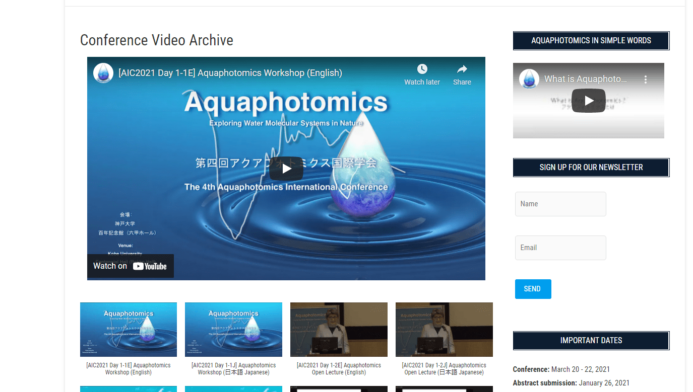
Finally, a Christmas will not be Christmas without presents. And as our Christmas present, we decided to make all the lectures from The 4th International Aquaphotomics Conference free for public. You can refresh your memories of how it all went in March, by watching a short digest video and find all the lectures and workshop on our Aquaphotomics YouTube channel.
We are looking forward to seeing you at the Aquaphotomics Christmas Special!
We are very pleased to announce that the video archive for “the 4th Aquaphotomics International Conference” is now available for free on the conference website.
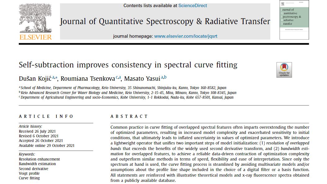
We are very pleased to announce that a new article on Aquaphotomics has been accepted and will be published in the “Journal of Quantitative Spectroscopy and Radiative Transfer” (Volume 277, January 2022, 107991).
“Self-subtraction improves consistency in spectral curve fitting” by Dušan Kojić, Roumiana Tsenkova, and Masato Yasui
Highlights • A purely functional algorithm for resolving positions and widths of overlapped bands is presented on the basis of self-subtracted spectra. • Significant improvements, demonstrated both theoretically and practically, over the popular second derivative transform are achieved. • The method eliminates any bias related to the choice of a basis function because it outputs the least-squares fit to the data. • Higher consistency with retained accuracy follow naturally from a systematic analysis of XRF spectra, across a variety of Voigt profile line shapes.
The end of this year will mark an important step in aquaphotomics development on a global level. The first aquaphotomics book titled “Aquaphotomics for Bio-diagnostics in Dairy – Applications of Near-Infrared Spectroscopy” is scheduled to be published by Springer on December 11, 2021.
The book is a result of more than 20 years of experience of working with near infrared spectroscopy in dairy and describes applications for measurement of raw milk components like fat, protein, lactose, urea and fatty acids. Further, it investigates factors that influence the accuracy of measurements like individuality of the dairy cow and then, for the first time, proceeds to establish the near infrared spectroscopy as a disease and bio diagnostics tool. The book shows that near infrared spectroscopy can be used for measurement of somatic cell count, mastitis diagnosis, estrus detection and other parameters of health and nutrition status of the animals thanks to the use of usually neglected water absorbance bands. These results present near infrared spectroscopy as a tool that provides a valuable feedback for dairy farm management and optimization of health status of animals.
The book shows how the utilization of water absorption regions in near infrared spectra brought entirely new dimension to NIRS which led to recognition of informational value of water spectra and led to the development of aquaphotomics, a novel science whose foundation was exactly the dairy research work. With more than 20 chapters, describing in detail experimental work, the book offers easy replication of performed experiments and can serve as a guide for anyone interested in application of aquaphotomics for bio-diagnostics.
The release of the book is schedule for 11th December, and can be found at the Publisher website page https://www.springer.com/gp/book/9789811671135. Since the publisher website is going to have a makeover in November, if the above provided link is not available, feel free to contact us for more information. Or you can preorder the book using the following links:
For all interested in aquaphotomics research and looking how to explain it to someone in simple words, we would like here to share an excellent short article, published at the beginning of this year as a part of news in Chemistry Research. The article Using light to reveal water impurities can be found at the following link:
The past months have been very productive in terms of publications for the Aquaphotomics Team at Kobe University. The latest paper, written in collaboration with a group from Keio University in Tokyo and Peking University in Health Science Center, Beijing, is about one of the very common topics – discrimination between different waters. The article is published on Oct 12th, in Applied Science Journal as a part of the special issue Novel Spectroscopy Application in Food Detection.
Behind this article is an interesting story of a special milestone. The paper took few years of working and several rejections, in the end being but forgotten and overshadowed by topics that were seemingly more popular. And, then recently, due to a set of circumstances the paper came up in conversation and it was decided that a bit more attention should be given to the patient, waiting manuscript.
And then, a funny thing happened during publication. One of the received reviews started with almost a dash of boredom, asking (to paraphrase) “we already know that water spectral patterns can be used for this …and the temperature perturbation influence is also well-known …so what exactly is new here?”
That was it – the milestone. Some things had changed. Aquaphotomics has become mainstream. It was a pleasant discovery!
So, what is really new in this paper?
First novelty can be immediately spotted in the title – Aquaphotomics Reveals Subtle Differences between Natural Mineral, Processed and Aged Water Using Temperature Perturbation Near-Infrared Spectroscopy. This is the first publication that examined the “processed and aged waters”. The “processed” water can be a broad category which in later years is receiving more and more attention due to the often-protected trade secrets do not reveal how the water is really treated to achieve a claimed functionality. Usually, traditional ways of analysis using physical and chemical measurements fail to return any significant conclusions and support the claims of some health benefits. Despite, the market is flooded with such water products. On the other hand, there is a popular belief that the bottled water usually has no expiry date. That it does not change at all. The quality standards do not address this issue, and it is very difficult to even express what the quality parameters are in such case.
The newly published paper presents exactly this case, which actually very often appears in our research practice. It deals with the discrimination of water spectra, when it extremely difficult, in most of the cases even impossible to find the differences or change in the spectra of waters after processing or after some period of storage, when using traditional ways of multivariate analysis.
In the paper it was shown that solution to the problem is sample perturbation by temperature and using water spectral pattern (WASP) to track how water systems evolve with perturbation; no matter how small and subtle the differences were in the beginning, they will be revealed through the dynamics of WASPs. With this, the paper proposes a perturbation protocol for characterization of waters as a solution to difficult discrimination problems, and expresses hopes that these new findings would also be considered by developers of optical measurement systems, where it would be sufficient to just enable perturbation by consecutive light irradiation, it doesn’t even have to be temperature.
The freshly published paper can be found at the following links.
C.Malegori, J. Muncan, E.Mustorgi, R.Tsenkova, P. Oliveri, Analysing the water spectral pattern by near-infrared spectroscopy and chemometrics as a dynamic multidimensional biomarker in preservation: rice germ storage monitoring, Spectrochimica Acta Part A: Molecular and Biomolecular Spectroscopy, Volume 265, 2022, 120396
It is our special pleasure to announce the publication of a new research paper by Aquaphotomics Research Department. The article written in collaboration with our Italian colleagues, titled “Analyzing the Water Spectral Pattern by Near-Infrared Spectroscopy and Chemometrics as a Dynamic Multidimensional Biomarker in Preservation: Rice Germ Storage Monitoring“, has been published in Spectrochimica Acta Part A: Molecular and Biomolecular Spectroscopy on 14th September.
The words “special pleasure” are insufficient to describe how we actually feel seeing this work published after two and more years of immense work. Precisely year ago, the last week of September we were in the midst of the series of experiments, with the laboratory cramped with all kinds of food, working from dawn to dusk, measuring water activity and the near infrared spectra. All that, just to be able to confirm that what we saw in the spectral data of rice germ during storage is a part of a universal phenomenon.
The results published in this paper are a first step in the series of articles that will follow which tackle this mystery of water activity and what it means to be alive, to be preserved, what exactly is deterioration and why things fall apart. And yes, of course, the answer is in the water. Not the quantity, but the molecular structure.
The Rice germ monitoring study provides the first comprehensive analysis of the state of water as a function of time and the initial water activity during storage. Using non-destructive, near infrared spectroscopy, advanced methods of data analysis (chemometrics) and a novel, aquaphotomics findings about the water structure-functionality relationship, we were able to describe the changes during storage only through the analysis of modifications in water molecular structure presented as spectral pattern of rice germ.
Our special gratitude goes to the leading authors Cristina Malegori, Paolo Oliveri and Eleonora Mustorgi who performed the experiments, masterfully executed chemometrics analysis and were kind to share their data and findings with us in order to pursue this research story. It contributed to our personal (especially in the patience section) and aquaphotomics growth immensely.
We hope this article will inspire similar experimental and data analysis studies of food and other products that will clarify the role of water in biological structures and their preservation, resulting in more efficient processing and preservation strategies.
The article can be found at the links below. Enjoy and share the water activity story!
Aquaphotomics group of Biomeasurement Technology Laboratory at Kobe University, Japan, has started functioning as Aquaphotomics Research Department since April this year.
Our first publication, under the new name is opening a new venue of using Aquaphotomics for cell development studies. Therefore it deserves a somewhat special treatment. The article “Water Spectral Patterns Reveals Similarities and Differences in Rice Germination and Induced Degenerated Callus Development” has been published in MDPI’s Journal Plants few days ago, and can be downloaded at the links provided below.
The paper is a part of the Special Issue Seed Physiology focused on the latest findings in this research area. The topic may seem mundane and ordinary, but translated into the everyday language we could say the title of the paper is “what is wrong with this rice?” and “can I predict if this rice will grow normally or not?”. If only thing that we worry about is how to produce rice, it may not seem much. But, as the guest editor of the special issue nicely put it “Plants start out their life as a seed” – to look at seed is to look at the origin of life.
In the paper, we found out what happens in the seed that determines if it will develop normally into a nice rice plant, or if it will degenerate and form callus. The collaborative effort of aquaphotomics groups in Hungary and Japan, resulted in finding that the healthy seeds, despite many individual differences, will go through orchestrated phases in water structuring and restructuring during normal growth. On the other hand, the degenerated growth showed random changes in water structure, without any specific patterns, without phases, without common characteristics.
The promising results of SWNIR spectroscopy coupled with aquaphotomics suggest the strong potential of the technique for rice seed authentication and characterization, and beyond that, for detection of abnormalities in cell growth and development, which may offer excellent feedback for variety of early warning systems in different areas of research.
Peanuts are one of the most common allergens, causing very severe allergic reactions. Peanut allergy accounts for 59% of the total number of food allergies and affects approximately 3 in every 100 children. The exact cause of allergy is not clear, but at least 11 specific proteins are recognized as peanut allergens. The allergen proteins can be made hypoallergenic after heating, which is called desensitization treatment. Essentially, this treatment can make modifications in protein structure, and the body no longer recognizes it as a pathogen, thus avoiding the allergic reaction.
The research group of Prof. Hengchang Zang at School of Pharmaceutical Sciences, Shandong University is dedicated to unraveling the relationship between the structure of proteins and their functionality, using aquaphotomics. Among many other biomolecules, this laboratory also explored the changes of the peanut allergen during heating process. In a recent publication, “Research on the Structure of Peanut Allergen Protein Ara h1 Based on Aquaphotomics” they applied aquaphotomics, using water as the probe to explain the detailed structural changes of peanut allergen protein Ara h1 during the heating process.
The first author of the article, published in June issue of Frontiers in Nutrition, is young Miss Mengqi Zhang, who is interested in applying Aquaphotomics to understand the structural information of natural products that could affect its function.
Aquaphotomics processing tools including principal component analysis (PCA), continuous wavelet transform(CWT), and two-dimensional correlation spectroscopy (2D-COS) were utilized for better understanding the thermodynamic changes, secondary structure, and the hydrogen bond network of Ara h1. The results showed that about 55 oC could be a key temperature which was the structural change point. During the heating process, the hydrogen bond network was destroyed, free water was increased, and the content of the protein secondary structure was changed. This discovery revealed the interaction between the water and Ara h1 from the perspective of water molecules and explained the effect of temperature on the Ara h1 structure and hydrogen-bonding system. Thus, it described a new way to explore the thermodynamic properties of Ara h1 from the perspective of spectroscopy and laid a theoretical foundation for the application of temperature-desensitized protein products.
The research was published June 18, 2021 in Frontiers of Nutrition, as a part of the research topic The Future Food Analysis. (https://doi.org/10.3389/fnut.2021.696355)
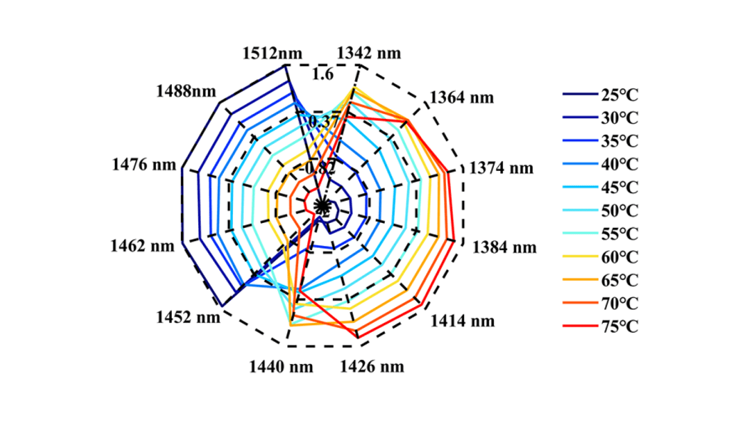
This research showed that during the heating process, the water spectra of the Ara h1 aqueous solution gradually shifted from high wavelength to low wavelength (Figure 1). The structures of Ara h1 and its spectra were changed under the influence of temperature. During the heating process, the strong hydrogen bonds were destroyed gradually, weak hydrogen bonds and other structures were formed. Under the influence of this effect, the water molecules were bounded by weak hydrogen bonds increasing. As the temperature rose again, the weak hydrogen bonds were also destroyed, the absorbance was weakened there, and the free water structure was formed. When the temperature was below 55oC, the WAMACS of the aquagram was mainly biased towards high wavelengths. At this time, the hydrogen bond network structure of Ara h1 aqueous solution was stable, and the hydration was strong between water and the protein surface, which had little effect on the protein structure. When the temperature was higher than 55oC, the aquagram tended to be at low wavelengths, the hydrogen bond network was broken, hydration was weakened, the structure of Ara h1 aqueous solution was greatly changed, and the protein precipitated and aggregated, and the hydrophobicity of the protein increased. It led to the decrease of sensitization ability.
Figure 1 The aquagram of Ara h1
The structures of Ara h1 had undergone major changes at around 55 oC, causing a rapid increase in the β-sheet content of the amide A /III (Figure 2 and Table 1). The α-helical content had been rising during the heating process. The secondary structures of the protein changed, and the side chain structures of the protein were broken, resulting in corresponding decreases of the sensitization ability of Ara h1.
Figure 2 Transformed spectra calculated by CWT of Ara h1 aqueous solution (2050~2350nm) (A); Absorbance of the peaks at 2183 nm (B), 2210 nm (C), 2288 nm (D) in the original spectra of Ara h1 aqueous solution during the heating process.
Table 1 Characteristic absorption of Ara h1 aqueous solution in NIR spectra
number
Wavelength (nm)
characteristic absorption band
1
2060
the N-H bending vibration; the second overtone of an -OH bending vibration of water
2
2183
Amide B / II
3
2210
β-fold
4
2288
α- helix
5
2342
-CH2 side chain
A method for characterizing the regularity of protein structural changes without labeling was established through this research. Compared with the conventional analysis method for structural changes, it was easy to operate and had high sensitivity. At the same time, it was based on the interaction between the allergen protein Ara h1 and the water structure, revealed the temperature point of the Ara h1 protein structural changes. It laid a theoretical foundation for food processing technology, and also provided a new idea to explore the interaction of various molecules in the life system.
For more information about this research, please contact the corresponding authors.
Mengqi Zhang
First Author
School of Pharmaceutical Sciences, Cheeloo College of Medicine, Shandong University, Jinan, China
Hengchang Zang
Corresponding author
School of Pharmaceutical Sciences, Shandong University, Jinan, China