Aquaphotomics group of Biomeasurement Technology Laboratory at Kobe University, Japan, has started functioning as Aquaphotomics Research Department since April this year.
Our first publication, under the new name is opening a new venue of using Aquaphotomics for cell development studies. Therefore it deserves a somewhat special treatment. The article “Water Spectral Patterns Reveals Similarities and Differences in Rice Germination and Induced Degenerated Callus Development” has been published in MDPI’s Journal Plants few days ago, and can be downloaded at the links provided below.
The paper is a part of the Special Issue Seed Physiology focused on the latest findings in this research area. The topic may seem mundane and ordinary, but translated into the everyday language we could say the title of the paper is “what is wrong with this rice?” and “can I predict if this rice will grow normally or not?”. If only thing that we worry about is how to produce rice, it may not seem much. But, as the guest editor of the special issue nicely put it “Plants start out their life as a seed” – to look at seed is to look at the origin of life.
In the paper, we found out what happens in the seed that determines if it will develop normally into a nice rice plant, or if it will degenerate and form callus. The collaborative effort of aquaphotomics groups in Hungary and Japan, resulted in finding that the healthy seeds, despite many individual differences, will go through orchestrated phases in water structuring and restructuring during normal growth. On the other hand, the degenerated growth showed random changes in water structure, without any specific patterns, without phases, without common characteristics.
The promising results of SWNIR spectroscopy coupled with aquaphotomics suggest the strong potential of the technique for rice seed authentication and characterization, and beyond that, for detection of abnormalities in cell growth and development, which may offer excellent feedback for variety of early warning systems in different areas of research.
Peanuts are one of the most common allergens, causing very severe allergic reactions. Peanut allergy accounts for 59% of the total number of food allergies and affects approximately 3 in every 100 children. The exact cause of allergy is not clear, but at least 11 specific proteins are recognized as peanut allergens. The allergen proteins can be made hypoallergenic after heating, which is called desensitization treatment. Essentially, this treatment can make modifications in protein structure, and the body no longer recognizes it as a pathogen, thus avoiding the allergic reaction.
The research group of Prof. Hengchang Zang at School of Pharmaceutical Sciences, Shandong University is dedicated to unraveling the relationship between the structure of proteins and their functionality, using aquaphotomics. Among many other biomolecules, this laboratory also explored the changes of the peanut allergen during heating process. In a recent publication, “Research on the Structure of Peanut Allergen Protein Ara h1 Based on Aquaphotomics” they applied aquaphotomics, using water as the probe to explain the detailed structural changes of peanut allergen protein Ara h1 during the heating process.
The first author of the article, published in June issue of Frontiers in Nutrition, is young Miss Mengqi Zhang, who is interested in applying Aquaphotomics to understand the structural information of natural products that could affect its function.
Aquaphotomics processing tools including principal component analysis (PCA), continuous wavelet transform(CWT), and two-dimensional correlation spectroscopy (2D-COS) were utilized for better understanding the thermodynamic changes, secondary structure, and the hydrogen bond network of Ara h1. The results showed that about 55 oC could be a key temperature which was the structural change point. During the heating process, the hydrogen bond network was destroyed, free water was increased, and the content of the protein secondary structure was changed. This discovery revealed the interaction between the water and Ara h1 from the perspective of water molecules and explained the effect of temperature on the Ara h1 structure and hydrogen-bonding system. Thus, it described a new way to explore the thermodynamic properties of Ara h1 from the perspective of spectroscopy and laid a theoretical foundation for the application of temperature-desensitized protein products.
The research was published June 18, 2021 in Frontiers of Nutrition, as a part of the research topic The Future Food Analysis. (https://doi.org/10.3389/fnut.2021.696355)
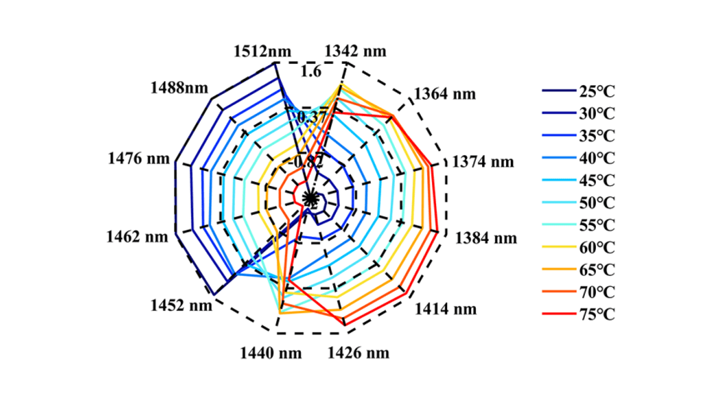
This research showed that during the heating process, the water spectra of the Ara h1 aqueous solution gradually shifted from high wavelength to low wavelength (Figure 1). The structures of Ara h1 and its spectra were changed under the influence of temperature. During the heating process, the strong hydrogen bonds were destroyed gradually, weak hydrogen bonds and other structures were formed. Under the influence of this effect, the water molecules were bounded by weak hydrogen bonds increasing. As the temperature rose again, the weak hydrogen bonds were also destroyed, the absorbance was weakened there, and the free water structure was formed. When the temperature was below 55oC, the WAMACS of the aquagram was mainly biased towards high wavelengths. At this time, the hydrogen bond network structure of Ara h1 aqueous solution was stable, and the hydration was strong between water and the protein surface, which had little effect on the protein structure. When the temperature was higher than 55oC, the aquagram tended to be at low wavelengths, the hydrogen bond network was broken, hydration was weakened, the structure of Ara h1 aqueous solution was greatly changed, and the protein precipitated and aggregated, and the hydrophobicity of the protein increased. It led to the decrease of sensitization ability.
Figure 1 The aquagram of Ara h1
The structures of Ara h1 had undergone major changes at around 55 oC, causing a rapid increase in the β-sheet content of the amide A /III (Figure 2 and Table 1). The α-helical content had been rising during the heating process. The secondary structures of the protein changed, and the side chain structures of the protein were broken, resulting in corresponding decreases of the sensitization ability of Ara h1.
Figure 2 Transformed spectra calculated by CWT of Ara h1 aqueous solution (2050~2350nm) (A); Absorbance of the peaks at 2183 nm (B), 2210 nm (C), 2288 nm (D) in the original spectra of Ara h1 aqueous solution during the heating process.
Table 1 Characteristic absorption of Ara h1 aqueous solution in NIR spectra
number
Wavelength (nm)
characteristic absorption band
1
2060
the N-H bending vibration; the second overtone of an -OH bending vibration of water
2
2183
Amide B / II
3
2210
β-fold
4
2288
α- helix
5
2342
-CH2 side chain
A method for characterizing the regularity of protein structural changes without labeling was established through this research. Compared with the conventional analysis method for structural changes, it was easy to operate and had high sensitivity. At the same time, it was based on the interaction between the allergen protein Ara h1 and the water structure, revealed the temperature point of the Ara h1 protein structural changes. It laid a theoretical foundation for food processing technology, and also provided a new idea to explore the interaction of various molecules in the life system.
For more information about this research, please contact the corresponding authors.
Mengqi Zhang
First Author
School of Pharmaceutical Sciences, Cheeloo College of Medicine, Shandong University, Jinan, China
Hengchang Zang
Corresponding author
School of Pharmaceutical Sciences, Shandong University, Jinan, China
NIRS is a modern analytical technique that makes use of the material information contained in the near infrared spectrum region and relies on chemometrics and computer technology to carry out qualitative and quantitative analysis of organic substances. It is one of the most important tools in PAT technology. NIRS has become one of the most rapidly developed new analytical techniques in the past decade due to its advantages of fast analysis speed, no pretreatment, no pollution, and simultaneous analysis of multiple components.
Drug quality and its uniformity is one of the important basic characteristics of drugs, and it is the core of drug efficacy and drug risk management. Many advanced pharmaceutical companies have adopted NIR spectroscopy for quality analysis and control of the whole process. However, the application of NIR spectroscopy in the pharmaceutical field is still in the research stage, and the common problems of many methodologies need to be further studied and understood. It is necessary to conduct theoretical discussion and experimental research on how to integrate the implementation of engineering, drug registration regulations, and the restrictions of GMP clauses. The consistency of drug efficacy and the improvement of drug re-registration standards all need the support of NIR technology. Therefore, in recent years, our research group has been devoted to the theoretical exploration and application research of the application of NIR spectroscopy in the pharmaceutical field.
Aquaphotomics related work
Hydration plays an important role in the stability of protein structure and function. The dynamic characteristics of hydration play a key role in biochemical processes, including protein folding, enzyme function, and molecular recognition. Protein purification is affected by hydration and dehydration. We used NIR spectroscopy to study the morphological changes of the hydration layer around protein molecules and detected the network structure of hydrogen bond water around protein molecules by changing the protein concentration, thus providing theoretical support for the stability of protein structure.
We are happy to say that our Conference is still the talk of the town!
You can read about it in the latest article “The 4th Aquaphotomics International Conference – Report ” published in Volume 32, Issue 3-4 of NIR news. We are always glad to be a part of the NIR news.
Jean-Michel Roger (left), Alexander Mallet (right)
It is a pleasure to announce the recent publications which could be of immense importance for aquaphotomics researchers. Both publications originate from the group of Jean-Michel Roger, (INRAE, UMR ITAP, Montpellier University and ChemHouse Research Group, France) one of the world experts in preprocessing and analysis of spectral data. The first author on both publications is Alexander Mallet, a young PhD researcher, who is working on revealing both physical and chemical effects of the water on the near infrared spectra, and who spent few months in the Biomeasurement Technology Laboratory, of Prof. Dr. Tsenkova at Kobe University, last year. The works are of particular importance for correct spectroscopic assignments, correct modeling and interpretation of the structure of water.
The first publication describes the results of the dynamic study of near-infrared spectra acquired during a drying process with a focus on the first overtone OH absorbance region:
The second one continues to analyze and compare moisture content effects in one comprehensive experiment with a wide variety of biochemical and physical types in order to understand water effects and how they relate to the substrate properties.
Both publications are heartily recommended and we expect to see their influence on aquaphotomics development. Most sincere congratulations to French group from the Aquaphotomics Research Department in Kobe. We are eagerly awaiting more.
Dear Conference Attendees, Friends, and Colleagues,
I would like to express my deepest gratitude for your invaluable contribution to the 4th Aquaphotomics International Conference held in March 2021 at Kobe University, Japan.
The conference was held both online and onsite on the first day, and was online only on the second and third day from the perspective of preventing the spread of COVID-19.
The conference was well attended with 248 participants from 23 countries (including 70 participants who attended the open lecture only) that greatly exceeded the 114 participants in the 2018 Symposium.
Thank you very much again for making this conference a great success! We will continue to pursue our research activities at the Aquaphotomics International Society.
Yours sincerely,
Roumiana Tsenkova Chairperson, Aquaphotomics International Society
We are very pleased to announce that a new article on Aquaphotomics has been published in “Sensors” (Volume 21, Issue 1, December 2020).
“Real-Time Monitoring of Yogurt Fermentation Process by Aquaphotomics Near-Infrared Spectroscopy” by Jelena Muncan, Kyoko Tei, Roumiana Tsenkova.
“Automated quality control could have a substantial economic impact on the dairy industry. At present, monitoring of yogurt production is performed by sampling for microbiological and physicochemical measurements. In this study, Near-Infrared Spectroscopy (NIRS) is proposed for non-invasive automated control of yogurt production and better understanding of lactic acid bacteria (LAB) fermentation. UHT (ultra-high temperature) sterilized milk was inoculated with Bulgarian yogurt and placed into a quartz cuvette (1 mm pathlength) and test-tubes. Yogurt absorbance spectra (830–2500 nm) were acquired every 15 min, and pH, in the respective test-tubes, was measured every 30 min, during 8 h of fermentation. Spectral data showed substantial baseline and slope changes with acidification. These variations corresponded to respective features of the microbiological growth curve showing water structural changes, protein denaturation, and coagulation of milk. Moving Window Principal Component Analysis (MWPCA) was applied in the spectral range of 954–1880 nm to detect absorbance bands where most variations in the loading curves were caused by LAB fermentation. Characteristic wavelength regions related to the observed physical and multiple chemical changes were identified. The results proved that NIRS is a valuable tool for real-time monitoring and better understanding of the yogurt fermentation process.”
We are very pleased to announce that a new article on Aquaphotomics has been published in “Food Control” (Volume 122, April 2021, 107805).
“Near infrared aquaphotomics study on common dietary fatty acids in cow’s liquid, thawed milk” by Jelena Muncan, Zoltan Kovacs, Bernhard Pollner, Kentarou Ikuta, Yoshihisa Ohtani, Fuminori Terada, Roumiana Tsenkova.
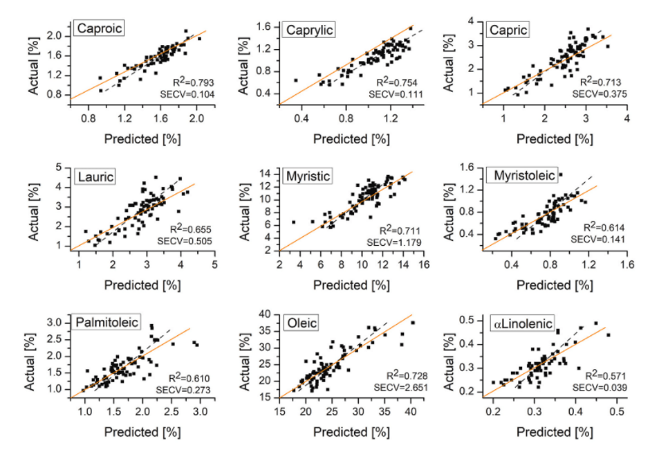
“This study was aimed at two objectives: 1) explore the possibility of near infrared (NIR) spectroscopy to determine the content of common dietary fatty acids (FAs) and fat content in cow’s liquid milk for better individual cow’s feed management; 2) using aquaphotomics obtain better understanding of interaction between water molecular structure and FAs and gain more insight into their functionality. For these purposes, 252 milk samples were collected from 9 cows during 14 weeks and FAs content measured using gas chromatography as a reference method. Applying aquaphotomics NIR spectral analysis, quantification was attempted using 1300–1850 nm region where both water and FAs absorbance bands are located. The results showed possibility of quantification for 8 common dietary FAs which was more accurate when spectral region was narrowed to 1600–1800 nm. In the external validation, good predictions (R2> 0.75, RPD>1.5), were obtained for caproic and caprylic and acceptable for capric, lauric, myristic, myristoleic, palmitoleic and oleic acid (0.55<R2<0.75, RPD>1.5). Interpretation of influential variables in regression models revealed contribution of water absorbance bands related to protonated and hydration water, which originate from the water-FAs interaction and are important for their self-organization into assemblies with different morphologies.”
Affiliation: Mississippi State University, Department of Biochemistry, Molecular Biology, Entomology, and Plant Pathology, Mississippi State, MS, USA 39762
Research Topics : Animal physiology, Amphibian conservation, NIR and NMR spectroscopy applied to animal and plant pathogens and diseases.
Description: Our research group started working with spectroscopic applications in animal physiology when Dr. Vance joined a research team focusing on NIR analysis of nutrition in the Giant Panda. We expanded our studies to develop NIR spectroscopic methodologies for determining basic physiological parameters (e.g. gender, reproductive status, age, disease) with the end goal of mapping the demographic movements of giant panda in-situ. Additionally, we have applied NIR spectroscopy to numerous other mammalian species (horses, cattle, elephants, okapi, leopards) and extended our research into non-mammalian taxa (anura, caudates, fish).
Aquaphotomics work: We have used Aquaphotomics to profile the reproductive status of Snow leopards and Amur leopards using NIR spectra collected from urine. In addition, we evaluated the reproductive cycling and performance of mares exposed to the fusarium mycotoxin Zearalenone, which causes hyperestogenism, by the analysis of blood serum spectra. Currently, we are using NIR spectroscopy and Aquaphotomics to analyze biochemical profiles of the pathogens Bovine Herpesvirus type 1, Bovine Respiratory Syncytial Virus, Mannhemia haemolytica, Xanthomonas spp, and Rhizoctonia solani. Our ultimate goal is to understand the biochemical changes occurring during the course of disease, and validate spectra profiling early stages of infection. Deterministic spectra feed into the development of NIR spectroscopy as a rapid, portable, non-destructive, and accurate diagnostic tool capable of reducing the time required for pathogen and disease detection and identification, which is a determining factor in infection-related mortality rates and the control of further disease spread.